Operates the platform
An agent that acts, not a chatbot that answers
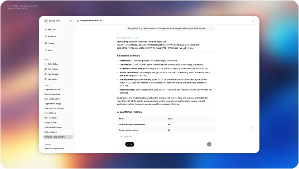
It searches the catalog, stages data, runs modules and sandboxed code, and publishes results through account-scoped, run-bound tools.
Agentic system for science
An agentic distributed system that runs real research where the data live.
A governed research agent that plans and acts through the platform's own services: it opens the files instruments produce, runs tools, code, and domain models against them, and keeps every result attached to the evidence that produced it. Not a chatbot beside the data — an agent that operates on it.
Operates the platform
An agent that acts, not a chatbot that answers
It searches the catalog, stages data, runs modules and sandboxed code, and publishes results through account-scoped, run-bound tools.
Governed autonomy
Bounded by measured confidence and hard limits
Sandboxed execution, independent runaway guards, per-result confidence, and a trust boundary the agent cannot rewrite.
The Intelligence Loop
Corrections become safely promoted models
Reviewer feedback drives gated finetuning, a deterministic canary, and instant rollback — implemented, not illustrated.
Measured, not promised
Frontier-tier responsiveness, self-hosted
59 ms to first token and 263 tokens per second on hardware the lab controls, with zero errors under load.
Collaborators
Demonstrated today
Governed autonomy, uncertainty-bounded reasoning, and a closed human-in-the-loop cycle are not a roadmap here. They run on a five-node production cluster. These numbers were measured live, end to end, with zero errors across every owned-tier ramp.
59 ms
Median time to first token from the self-hosted coordinator model.
263 tok/s sustained
~2 ms
Native OME-Zarr metadata on terabyte-scale stores. Cold equals warm.
~80 MB resident
0 errors
Across every owned-tier load ramp, and every real run in a 30-minute live observation.
zero failed runs
51,625 rps
Static delivery at the edge; roughly 39,000 rps of authenticated API with headroom.
CPU-bound, not saturated
10,000+
Concurrent browsing users in the derived envelope, with dozens of simultaneous agent runs.
measured knees
4.7 GB → 46 MB
A gigapixel image read, bounded after the sensor-data hardening pass.
fails closed on malformed input
Responsiveness is a deployment property, not a claim to out-reason a frontier flagship: the model is co-located with the data, so there is no wide-area hop. Full method, tables, and the third-party output-speed comparison are in the performance envelope.
The Lens viewer
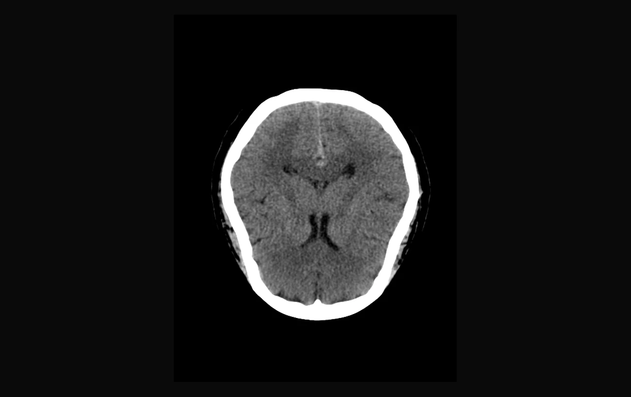
Lens is where the workbench meets the data. It opens the multidimensional files scientific instruments actually write, with real physical units and windowing, and it renders the figures the agent generates in the same surface — so a result never leaves the context that produced it.
Clinical CT · NIfTI
The instrument's data, with its physics intact.
A thoracic-window head CT with Hounsfield-unit windowing, z-scrub across 34 planes, and anisotropic voxel spacing read straight from the volume — not flattened to a screenshot.
Agent output · detection density
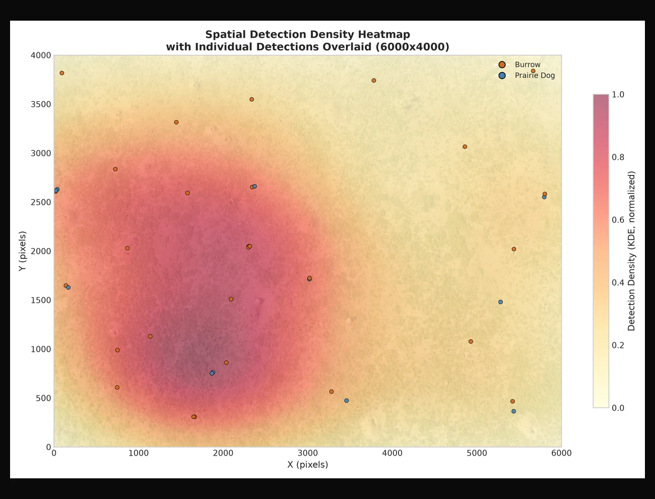
The agent's result, in the same surface.
A spatial detection-density heatmap over a 6,000 × 4,000 aerial survey, with individual prairie-dog and burrow detections overlaid — an artifact the agent produced during a run, opened like any other resource.
One scientific record
A run is not a single request and response. It is staging, inspection, tool selection, code execution, model calls, generated figures, and review — carried out where the data live and kept intact through the ordinary failures of browsers, workers, and services.
Upload directly or connect a BisQue resource with metadata, dimensions, annotations, and collection context intact.
It reasons over the request, stages named data, and runs specialized tools, code, vision services, and workers against the selected resources.
Execution is sandboxed and durably checkpointed; a worker restart resumes from the last completed step instead of repeating the work.
Return to the report, figures, tables, metrics, run events, and downloadable artifacts without reconstructing the experiment from memory.
Governed autonomy
We concede the premise — language models hallucinate, and their outputs cannot be trusted unvalidated. So an agent that touches real data and real compute is bounded structurally, not asked nicely. Every claim below is enforced in code, and every action is recorded.
Sandboxed execution
No network, all Linux capabilities dropped, a read-only root filesystem, bounded CPU and memory, and a wall-clock ceiling the model itself cannot extend — from an image pinned by cryptographic digest and stamped into the run's provenance.
Bounded reasoning
A progress-stall detector, an idle watchdog, and a bounded completion cap halt trajectories that stop making progress. Every delegated result carries a machine-checked confidence level governed by an explicit decision rule.
Structural trust boundary
A run-bound worker can touch only the resources its own run authorized; the agent cannot alter its own skills or the organization's policy; and generated charts and workflows pass a reject-by-default validator before they render.
Current product
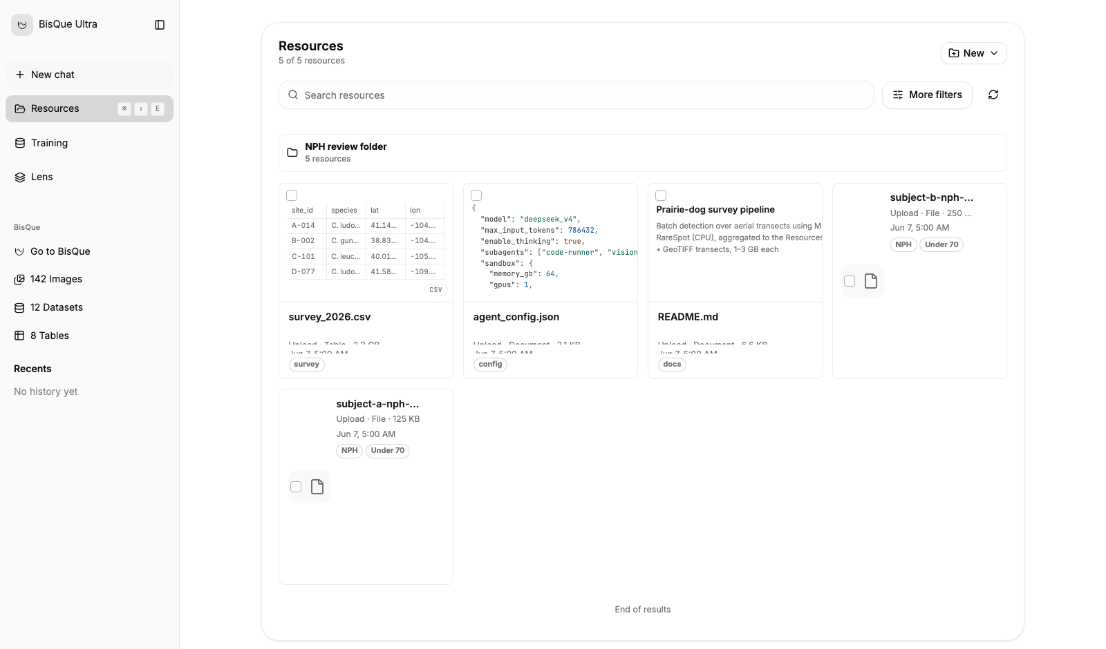
These are the current interfaces of the deployed system, each backed by the Go control plane and its durable contracts — the same frontend the local stack runs.
Resources
Search, filter, preview, organize, share, rename, restore, and move resources into analysis. The same system handles upload progress and recovery, collections, BisQue imports, direct viewers, and derived pyramids for large images — the catalog the agent searches and stages from.
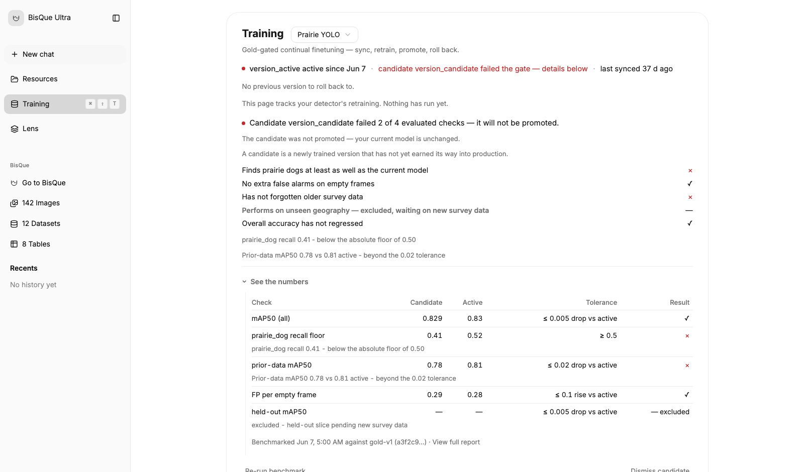
GoldGate training
Freeze a gold set, launch retraining, follow live progress, compare a candidate against the active model, and promote only when the declared gates pass. Canary routing and rollback keep the serving decision explicit after evaluation.
The Intelligence Loop
Domain models run where the data live and return results with quality flags. Confident results proceed with a full audit trail; low-confidence, high-value cases escalate to a targeted expert; validated corrections train the next model — safely. This is the loop BisQue Ultra runs, bounded at every step by measured confidence.
Bright 4B
The lab's flagship workload: a 4B-parameter foundation model that learns on the unit hypersphere to segment subcellular structures directly from 3D brightfield volumes. Native Sparse Attention, residual HyperConnections, soft Mixture-of-Experts, and anisotropic patch embedding address context, representation flow, adaptive capacity, and confocal geometry — and Ultra gives it a durable path from volume selection to figures, tables, and review.
Read the Bright 4B paper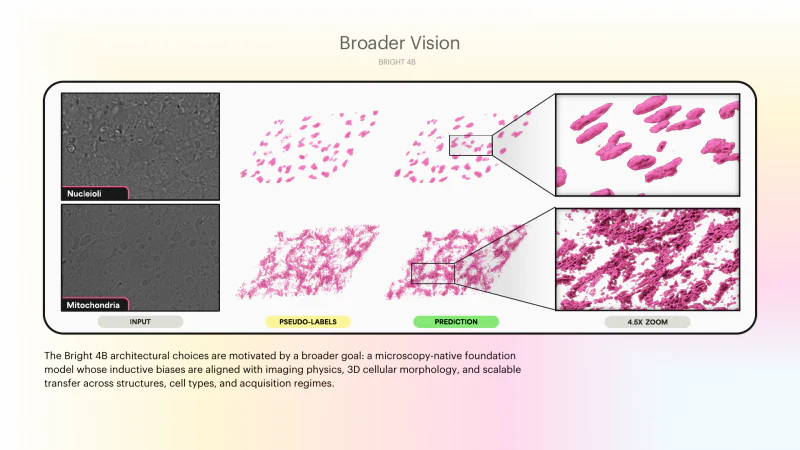
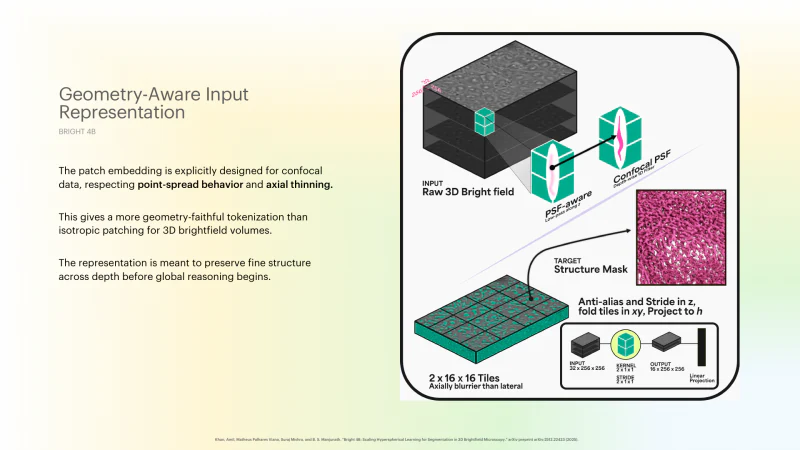
Representation
The model respects point-spread behavior and axial thinning before global reasoning begins, preserving fine structure across depth.
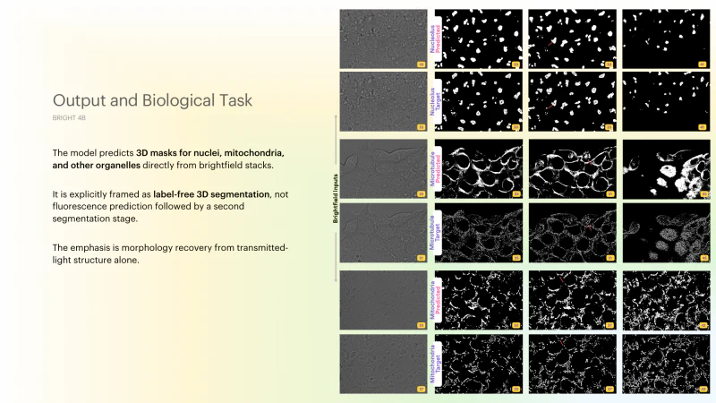
Biological output
Nuclei, mitochondria, microtubules, and related structures can be compared as segmentation outputs rather than treated as opaque model responses.
Published foundations
BisQue Ultra is not a wrapper around a chat model. It comes from a lab whose public work spans remote sensing, ecology, clinical neuroimaging, materials science, and bioimage analysis — each program shaped by the geometry, physics, or biology of its measurement, and each ending in code, data, or a platform path other researchers can inspect.
Hyperspectral remote sensing
Detects methane plumes from airborne hyperspectral imagery with a model designed around absorption wavelengths, plus the public Methane HotSpot dataset and BisQue visualization tooling.
CVPR 2023 Highlight
The repository includes source code, pretrained plume detection and segmentation weights, MHS dataset download scripts, and a BisQue online dataset viewer.
Ecological computer vision
Turns aerial imagery into detection, localization, and species-identification signals for wildlife monitoring, grounded in a verified dataset of 11k images and 28k annotations.
CVPR 2024
The public repository ships model code, pretrained detector weights, dataset tooling, and a BisQue visualization route for the Mara-Wildlife dataset.
Materials microscopy
Adapts super-resolution to EBSD orientation maps using crystallographic symmetry, quaternion-aware orientation recognition, and physics-aware losses.
npj Computational Materials 2022
The project connects network training, inference, IPF-map visualization, and BisQue module deployment for high-throughput materials characterization.
Clinical neuroimaging
Improves glioma segmentation by registering a brain parcellation atlas into each subject space and combining that location signal with multimodal MR images, 3D U-Net, DeepMedic, and ensemble learning.
Frontiers in Neuroscience 2020
The paper reports that location information improves patch-based neural networks for BraTS brain tumor segmentation and uses model diversity plus uncertainty reduction in a two-level ensemble.
Clinical neuroimage analysis
Segments regions of interest from CT brain scans, combines them with diffusion tractography and connectome features, and predicts Normal Pressure Hydrocephalus from measurable neuroanatomy.
BME Frontiers 2022
The article describes an automated NPH prediction method from CT scans that incorporates MRI diffusion tractography information, with reported gains over prior state-of-the-art precision and recall.
Bioimage analysis
Turns time-lapse 3D confocal image stacks into quantitative cell histories with rotation-equivariant 3D segmentation, adjacency-graph feature extraction, and graph-based tracking.
Scientific Reports 2023
The Scientific Reports paper states that code is available on GitHub and the method is available as a service through the BisQue portal.
Research program
Ultra Mode is BisQue Ultra's durable, typed multi-agent tier. Rather than ship it on a promise, we hold it to a falsifiable theory of when orchestration helps and a preregistered evaluation — and we publish the results that contradict us. It runs behind fail-closed admission while it is qualified.
Orchestration should win only with high coverage burden, decomposable work, compressible results, and an observable test — not from more agents or more thinking.
The model proposes a finite typed plan; the control plane validates ports, authorization, budget, and termination before any durable work runs. A plan proposal is never a queue command.
Seven arms isolate the mechanism; gates and margins are signed before an untouched lockbox is opened once. Inspecting an outcome and changing the system consumes it.
A generic planner did not beat a direct run; typed map-reduce took quality from 5-of-15 to 15-of-15; a model verifier accepted twelve wrong answers, so only a server seal commits success.
Open architecture
Data, run control, worker execution, model serving, and interface state are separate concerns on durable infrastructure. Postgres is the ordering authority; a partitioned NATS JetStream spine carries work with effectively-once semantics; the model layer stays replaceable.
Images, datasets, tables, metadata, annotations, modules, uploads, and collections remain the scientific substrate.
Auth, threads, runs, leases, training state, events, artifacts, and the OpenAPI contract sit on durable Postgres state, with multi-tenant isolation on every serving path.
JetStream dispatches long work to scalable workers that execute tools, code, paper workflows, and domain services, with idempotent redelivery.
The interface keeps resources, conversations, streamed steps, viewers, artifacts, training, and administration visible.
Evaluation questions
It plans and acts through the platform's own services. The agent searches a researcher's catalog, stages named data into its workspace, runs modules and code, calls domain models, and publishes results back through account-scoped, run-bound tools. Long trajectories are durably checkpointed and keyed to the run, so a worker restart resumes from the last completed step instead of re-executing the work.
Agent-generated code runs only inside a locked-down container with no network, dropped Linux capabilities, a read-only root filesystem, bounded CPU and memory, and a wall-clock ceiling the model cannot extend. Independent guards halt runaway loops, every delegated result carries a machine-checked confidence level, and the agent can read but never rewrite its own skills or the organization's policy.
Yes. The worker uses an OpenAI-compatible contract and can point at Ollama, vLLM, or another compatible server, self-hosted beside the data. The stable product contract is the data, tools, runs, artifacts, and evaluation logic; the model can change while the scientific record stays legible.
Yes. Docker Compose launches Postgres, NATS JetStream, the scientific image service and conversion worker, the Go control plane, a Deep Agents worker, and the React frontend. You provide a compatible model endpoint. An existing BisQue deployment is optional and adds shared images, datasets, metadata, and modules.
Ultra Mode — the durable, typed multi-agent orchestration tier — runs behind fail-closed admission while it is qualified against a preregistered evaluation. It is a research program with honest, published early results, not yet a public feature. The single-agent workbench described here is what runs today.
The materials surface is an evidence-gated research capability. Production promotion still requires designated live traces, ledger qualification, production sandbox and isolation evidence, external evaluation thresholds, and an attested promotion envelope.
Release notes
The launch brief gives the complete release view. The engineering and research notes go deeper on the performance envelope, sensor-data safety, model operations, and how we evaluate the orchestration tier.
Launch brief
The 2026.07 research release brings scientific file and viewer infrastructure, durable analysis, gold-gated training, and evidence-aware domain tools into one workbench.
July 13, 2026 · 2026.07First token in 59 ms, 263 tokens per second sustained, OME-Zarr metadata in two milliseconds, and zero errors across every owned-tier load ramp. The numbers behind an agent that runs where the data live.
When does orchestrating agents beat a single strong trajectory, and which mechanism causes the gain? A falsifiable theory, a typed workflow algebra, and a preregistered evaluation with honest negative results.
29 spec-correct stores across seven domains all render; 23 malformed stores all fail closed with precise errors; a gigapixel read that once cost 4.7 GB now costs 46 MB. The stress test that got us there.
A technical tour of the image service, conversion worker, resource model, and multidimensional viewers that let Ultra work with the files scientific instruments actually produce.
Why a scientific workbench needs more than a training button, and how GoldGate makes model versions, evaluation failures, canaries, and rollback visible.
Work with us
We are opening BisQue Ultra to research teams and frontier labs working with complex scientific data, domain models, autonomous computation, and reproducibility requirements. Tell us what you measure, what you need to run, and where the evidence currently breaks apart.